Our Research

The Wong Laboratory is in the Division of Neuropathology at the Johns Hopkins University School of Medicine. The overarching themes focus on the biology and pathobiology of an RNA splicing factor termed TDP-43 (TAR DNA/RNA binding protein 43kDa) that regulate the inclusion of cryptic exons, the loss of which underlies the pathogenic mechanism of several human age-related degenerative diseases, including Alzheimer’s Disease Related Dementia (ADRD), Amyotrophic Lateral Sclerosis (ALS), as well as Inclusion Body Myositis (IBM).  

Ongoing projects include:

  1. the development of in vitro and in vivo models of TDP-43 loss-of-function in the nervous system and skeletal muscle to reveal its biology in a cell- or organ-specific manner;
  2. the development of mouse models that reflect disease-specific mimics of ADRD, including AD-TDP and FTLD-TDP subtypes and IBM;
  3. testing of an AAV9 gene therapeutic strategy and the development of prognostic fluid biomarkers for these human diseases.

Below illustrates some of our recent works (click on each item to read more):

1. TDP-43 repression of nonconserved cryptic exons is compromised in ALS-FTD. (Science, 349(6248), 650-655, 2015)

Identification of TDP-43 associated cryptic exons in Homo sapiens.
(A) TDP-43 protein levels are greatly reduced when HeLa cells are treated with TDP-43 siRNA (*, nonspecific band). (B) Visualization of the cryptic exon located in EPB41L4A. Zoom in of gene annotation demonstrates that the cryptic exon (green arrow) resides in a non-conserved region. Strand specific analysis also verifies the incorporation of cryptic exons on the transcribing strand (fig. S12). (CF) IRF9 contains a transcriptional start site (C), KRT7 an exon extension (D), GPSM2 a standard cryptic exon (E), and INSR a polyadenylation site (F).

2. Depletion of TDP-43 decreases fibril and plaque β-amyloid and exacerbates neurodegeneration inanAlzheimer’s mouse model.

Forebrain depletion of TDP-43 increases relative levels of prefibril Aβ oligomers, while reducing Aβ plaques and fibril oligomers. Representative images of 4G8 stained plaques in AD amyloidosis model (AP) and AP mice lacking TDP-43 in forebrain neurons (cTAP)

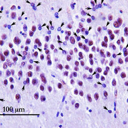
3. Cryptic exon incorporation occurs in Alzheimer’s brain lacking TDP-43 inclusion but exhibiting nuclear clearance of TDP-43. (Acta Neuropathologica 133:923-931, 2017)

Dentate gyrus region stained with TDP-43 phosphorylation-independent antibody. Arrows indicate neurons with nuclear clearance of TDP-43.

4. Splicing repression is a major function of TDP-43 in motor neurons.
(Acta Neuropathologica July 22, 2019; doi.org/10.1007/s00401-019-02042-8).

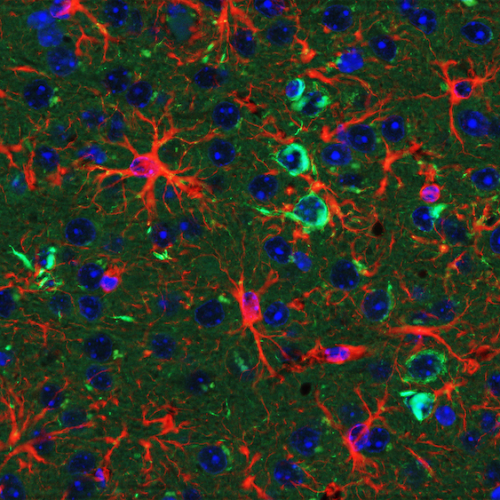
Immunostaining of p30 ChAT (red) and CTR (recognized by human-specific N-terminal TDP-43 antibody, green) in representative lumbar ventral horn sections of ChAT-IRES-Cre;TardbpF/+ and ChAT-IRES-Cre;TardbpF/F mice.

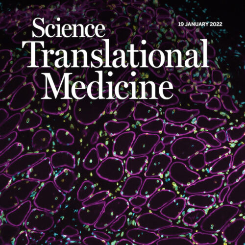
5. Loss of TDP-43 function and rimmed vacuoles persist after T cell depletion in a xenograft model of sporadic inclusion body myositis. (Science Translational Medicine 14, eabi9196, January 19, 2022)

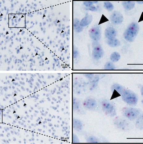
Cryptic Exon detection is a sensitive and specific assay for TDP-43 pathology in IBM biopsies. (A) Levels of TDP-43 are substantially reduced in myoblasts treated with TDP-43 siRNA (KD) compared to control siRNA (C). (B) Visualization of cryptic exons (green arrow) in myoblast cells with TDP-43 knockdown (MyoKD) compared to control (MyoC): ACOT11, SLC39AB, PFKP, and RHEBL1. (C) Immunohistochemical TDP-43 staining of muscle sections: control showing nuclear TDP-43 and IBM showing accumulation of TDP-43 in the cytoplasm (#) or nuclear clearing (arrows).