
Traumatic axonopathies after diffuse TBI: the role of the SARM1 (Wallerian) pathway and DLK (MAPK) cascade
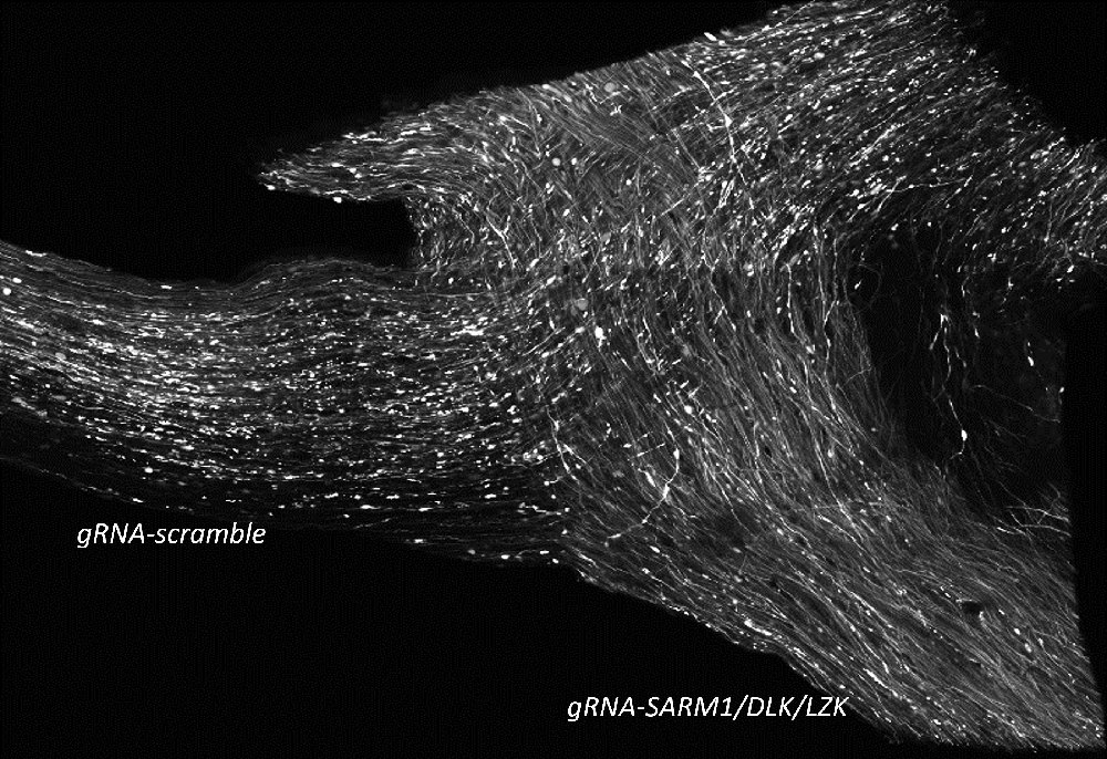
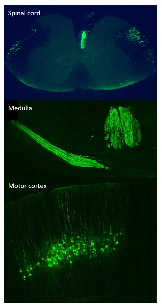
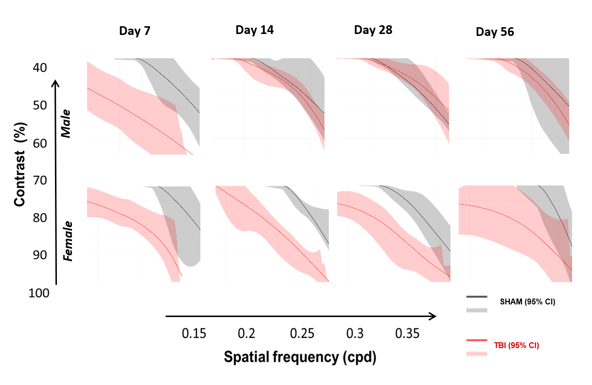
- NMNAT2-SARM1 signaling in traumatic axonopathy in the corticospinal and visual pathway: genetic (cre-lox, CRISPR) and pharmacological interventions for prevention and treatment of pathology and symptoms
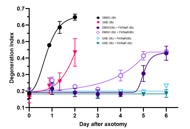
Characterization of the effects of small molecules acting on NAD biosynthetic pathways on the outcome of Wallerian degeneration: NAMPT inhibitors, DLK inhibitors, NaR alone and in combination
Characterization and biomarking of early stages of axonopathies
The role of axonal injury in accelerating tauopathy in animals harboring pathogenic tau mutants or seeded with Alzheimer tau prions
Vagus nerve stimulation-based neuromodulation for neuroinflammation in neurotrauma and related conditions
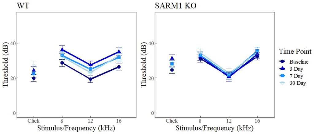
The role of SARM1 in traumatic injury of the auditory system
Neuropathology of contusional and diffuse axonal injury in the human brain
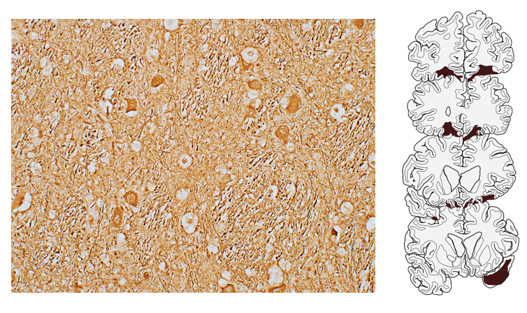
Effects of such focal injuries are localized to thalamus, but secondary effects of diffuse white matter injury are throughout gray matter.